Ultrasound Radiomics Enhances Kidney Disease Differentiation in T2DM Patients
A recent study highlights the effectiveness of ultrasound radiomics combined with machine learning in distinguishing diabetic kidney disease from non-diabetic kidney disease.
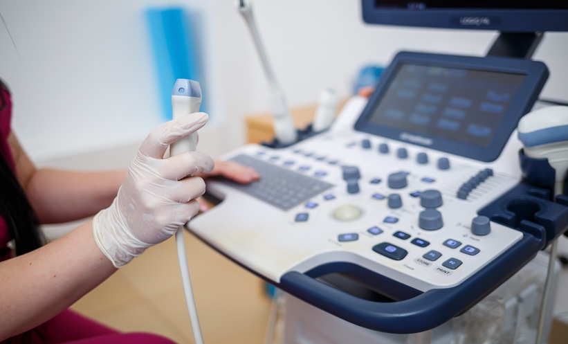
Ultrasound radiomics shows promise in differentiating DKD from NDKD in Type 2 diabetes patients.
The integrated XGBoost model achieved an impressive AUC of 0.991 in the training cohort.
Findings suggest potential for non-invasive preliminary screening in nephrology departments.
A multicentre retrospective study has revealed that ultrasound radiomics, when paired with machine learning, can effectively differentiate diabetic kidney disease (DKD) from non-diabetic kidney disease (NDKD) in individuals suffering from Type 2 diabetes mellitus (T2DM). This innovative approach offers a non-invasive method for initial disease differentiation, potentially transforming how kidney diseases are assessed and managed.
The research involved patients diagnosed with T2DM who underwent renal biopsy across three medical centers. The biopsy results were crucial in categorizing patients into DKD or NDKD groups, with any overlapping lesions classified under DKD. The study meticulously analyzed renal ultrasound images, focusing on specific regions to extract relevant radiomic features, while also employing statistical and machine learning techniques to identify clinical predictors.
Among the various machine learning models assessed, including support vector machines and random forests, the integrated XGBoost model stood out. It combined ultrasound radiomic data with estimated glomerular filtration rate (eGFR) and achieved an area under the curve (AUC) of 0.991 in the training cohort. Validation cohorts showed AUCs of 0.895 and 0.721, respectively, indicating variability in model performance. Additionally, the F1-scores reflected similar trends, showcasing the model's strengths and weaknesses across different cohorts.
The implications of this study are significant for clinical practice, particularly in primary healthcare settings and nephrology departments. The authors suggest that integrating ultrasound radiomics with existing clinical data could enhance the differentiation process between DKD and NDKD, aiding in early diagnosis and treatment decisions. This approach may also help streamline the decision-making process regarding the necessity of renal biopsies, potentially reducing patient risk and healthcare costs.
Despite the promising results, the study acknowledges that the performance of the model in external validation cohorts was less robust than in the training and internal validation cohorts. This highlights the need for further research to refine the model and ensure its reliability across diverse patient populations. Overall, the findings advocate for continued exploration of ultrasound radiomics and machine learning as valuable tools in the non-invasive differentiation of kidney diseases in T2DM patients.